
IGF-1 rs6218 polymorphisms modulate the susceptibility to age-related cataract
- Published
- Accepted
- Received
- Academic Editor
- Ramcés Falfán-Valencia
- Subject Areas
- Genetics, Genomics, Geriatrics, Ophthalmology
- Keywords
- Age-related cataract (ARC), Lens epithelial cells (LECs), Single nucleotide polymorphisms (SNPs), Insulin-like growth factor 1(IGF1), Apoptosis
- Copyright
- © 2024 Zou et al.
- Licence
- This is an open access article distributed under the terms of the Creative Commons Attribution License, which permits unrestricted use, distribution, reproduction and adaptation in any medium and for any purpose provided that it is properly attributed. For attribution, the original author(s), title, publication source (PeerJ) and either DOI or URL of the article must be cited.
- Cite this article
- 2024. IGF-1 rs6218 polymorphisms modulate the susceptibility to age-related cataract. PeerJ 12:e17220 https://doi.org/10.7717/peerj.17220
Abstract
Background
Single nucleotide polymorphisms (SNPs), as the most abundant form of DNA variation in the human genome, contribute to age-related cataracts (ARC) development. Apoptosis of lens epithelial cells (LECs) is closely related to ARC formation. Insulin-like growth factor 1 (IGF1) contributes to cell apoptosis regulation. Moreover, IGF1 was indicated to exhibit a close association with cataract formation. Afterward, an investigation was conducted to examine the correlation between polymorphisms in IGF1 and the susceptibility to ARC.
Methods
The present investigation was a case-control study. Venous blood draws were collected from the participants for DNA genotyping. Lens capsule samples were collected to detect mRNA and apoptosis. TaqMan RT-PCR was used to detect IGF1 polymorphism genotypes and qRT PCR was used to detect IGF1 mRNA levels in LECs. LEC apoptosis was evaluated through flow cytometry. The chi-square test was used to compare differences between ARCs and controls of each SNP.
Results
We found that the G allele frequency in the IGF1-rs6218 was higher in the ARCs than in the controls. Furthermore, it was observed that the rs6218 GG genotype exhibited a positive correlation to elevated levels of IGF1 mRNA in LECs. The IGF1 mRNA in the LECs and the apoptosis of LECs in nuclear type of ARCs (ARNC) was higher than the controls.
Conclusion
The susceptibility to ARC was related to IGF1-rs6218 polymorphism, and this polymorphism is associated with IGF1 expression at the mRNA level. Moreover, apoptosis in LECs of ARNCs was found to be increased.
Introduction
Age-related cataract (ARC) is the transformation of the crystalline lens from transparent to cloudy, it is a multifactorial disorder standing as the primary contributor to blindness globally, especially in middle-income and low-income countries (Cicinelli et al., 2023). Although surgery can restore vision in most patients, the procedure itself has some risks and complications, which is a heavy economic burden for patients in developing countries and may lead to surgical complications, even irreversible blindness (Bhandari & Chew, 2023; Karesvuo et al., 2022). The risk of ARC was found to be affected by several genetic and environmental factors, including oxidative damage and metabolic disorders, among others (Park & Lee, 2015; Tang et al., 2018). However, the definite pathogeny of ARC remains incompletely understood.
Genetic variations, especially single nucleotide polymorphisms (SNPs), participate significantly in ARC development, SNPs are the most abundant form of DNA variation in the human genome, they can exist at any location of a gene, incluing intron, coding and untranslated region (Zou et al., 2018). Some of our previous genes and SNPs were associated with ARC: LSS-rs2968 A allele might play a role in the formation and development of nuclear type of ARC risk (Zou et al., 2020); the NEIL2-rs4639 T allele was strongly associated with a protective role in ARCs (Kang et al., 2019); the XPC-rs2229090 C allele was associated with ARNC risk (Zou et al., 2018).
Lens epithelial cells (LECs) are the primary site of metabolic activity within the lens (Wang et al., 2017). Apoptosis of LECs is the cellular basis of cataract formation (Zheng et al., 2018). LECs apoptosis leads to loss of homeostasis and accumulation of crystallin proteins, which then leads to the formation of cataracts (Qi et al., 2022). Low expression of MSH3 leads to apoptosis of LECs, which may lead to the occurrence of ARC (Chen et al., 2022).
The insulin-like growth factor (IGF) system is involved to a great extent in governing multiple biological processes, including metabolic function, cellular development, cell proliferation, differentiation, and apoptosis (Tien et al., 2017; Willems et al., 2016). This IGF system comprises IGF-1/2 besides their cell surface receptors (IGF-1R/2R) and six specific IGF-binding proteins (IGFBPs), IGF-1R participates in gene expression regulation by forming transcriptional complexes, modifying the activity of chromatin remodeling proteins, and participating in DNA damage tolerance mechanisms. IGF-1 is a small protein that binds to IGFBPs in circulation and stimulates IGF-1R upon release, which undergoes self phosphorylation (Poreba & Durzynska, 2020; Cao et al., 2016). IGF1 can activate their receptors and trigger the phosphorylation of IGF1R itself and downstream signal transduction, including PI3K/AKT and Ras/Raf/ERK signal pathways, inhibit cell proliferation and promote cell apoptosis (Cao et al., 2020).
Study shows SNPs of IGF1R are related to eye diseases (Chiu et al., 2011; Cui et al., 2020). IGF1R-rs2872060 revealed a significant association with advanced AMD (Chiu et al., 2011), IGF1R-rs1546713 may affect susceptibility to ARCs (Cui et al., 2020). Moreover, IGF1 was indicated to exhibit a close association with cataract formation (Civil et al., 2000; Papier et al., 2022; Wang et al., 2022). IGF1 might decrease rats’ content of α-crystallin made in the lens Fibre cells, which leads to rat lens opacification and cataract formation (Civil et al., 2000). IGF1 promoted cataract formation might by promoting the epithelial-mesenchymal transformation of LECs (Wang et al., 2022). However, whether SNPs of IGF1 were related to ARC is still not clear.
Materials and Methods
Study participants
The study was granted approval from the Ethics Committee of Changzhou Third People’s Hospital (ethical approval number: 2021012) and followed the Declaration of Helsinki. The participants were aware of the study’s aim and subsequently provided their informed consent by signing the appropriate documentation.
All participants underwent a comprehensive ophthalmic assessment, which included examinations of visual acuity, lens using a slit lamp biomicroscope under transient and side illumination after mydriasis, and ophthalmoscopic. The classification of ARC based on the opacity region of the lens includes four subtypes: cortical cataract (CC), nuclear cataract (NC), posterior sub-capsular cataract (PSC), and mixed cataract (MC) (Klein, Klein & Linton, 1992). The diagnosis and grading of lens opacities were conducted following the Lens Opacities Classification System III (LOCS III) (Feng et al., 2022).
This study adopts a case-control design, involving cases and controls from a population based epidemiologic cohort of the Jiangsu Eye Study located in Jiangsu Eye Study in Qidong country. In addition, individuals who served as controls and were matched with the experimental terms of age and sex and had transparent lenses were selected from the same communities. The geographical region under investigation exhibits a relatively stable and ethnically homogenous population. The participants were individuals sharing no familial relationships and self-identified as belonging to the Han Chinese ethnic group. Consequently, 716 ARC patients (CC = 377, NC = 223, PSC = 48, MC = 68) and 685 controls were included (Table 1). The inclusion/exclusion details of the case-control design followed the previous study (Kang et al., 2019).
| Variable | n | Age (Mean ± SD) | p | sex | χ2 | p | |
|---|---|---|---|---|---|---|---|
| Male (%) | Female (%) | ||||||
| Controls | 685 | 69.54 ± 5.35 | 294 (42.92) | 391 (57.08) | |||
| ARCs | 716 | 69.59 ± 5.39 | 0.211 | 281 (39.25) | 435 (60.75) | 0.014 | 0.174 |
| C | 377 | 70.1 ± 5.64 | 0.451 | 167 (44.4) | 210 (55.6) | 0.052 | 0.524 |
| N | 223 | 68.9 ± 5.86 | 0.121 | 103 (46.4) | 120 (53.6) | 0.162 | 0.371 |
| PSC | 48 | 68.2 ± 4.16 | 0.192 | 22 (45.8) | 26 (54.2) | 0.026 | 0.491 |
| M | 68 | 70.1 ± 6.14 | 0.514 | 32 (47.1) | 36 (52.9) | 0.005 | 0.501 |
Note:
C, cortical cataract; N, nuclear cataract; PSC, posterior sub capsular cataract; M, mixed cataract.
In addition, we included additional 20 ARNC patients and 20 age-, sex- and ethnically matched controls from inpatients in our hospital from Jan 2019 to Dec 2021 (Table 2) to collect not only venous blood but also matched lens anterior capsule samples. Venous blood is used to detect its genotype, while lens anterior capsule samples were used to measure the mRNA levels of LECs and detect their apoptosis. The lens anterior capsule samples from ARC patients were obtained through anterior continuous circular capsulorhexis in phacoemulsification surgery. The lens anterior capsule samples from transparent lenses were acquired from patients who received lens extraction as part of vitrectomy procedures. The study excluded participants with lens trauma, diabetes, uveitis, glaucoma, and high myopia (>6D).
| Controls | ARNCs | ||||||||
|---|---|---|---|---|---|---|---|---|---|
| Samples | Age (y) | Sex | LOCSIII | Genotype | Samples | Age (y) | Sex | LOCSIII | Genotype |
| No. 1 | 65 | Female | N0C1P0 | AA | No. 1 | 58 | Female | N3C0P0 | GG |
| No. 2 | 57 | Male | N0C0P0 | AG | No. 2 | 58 | Female | N5C0P0 | AA |
| No. 3 | 53 | Female | N0C0P1 | AA | No. 3 | 75 | Male | N4C0P0 | AG |
| No. 4 | 65 | Female | N0C1P0 | GG | No. 4 | 75 | Male | N3C0P0 | AA |
| No. 5 | 57 | Male | N0C0P0 | AA | No. 5 | 55 | Male | N5C0P0 | AA |
| No. 6 | 73 | Male | N0C0P1 | AA | No. 6 | 72 | Male | N3C0P0 | AA |
| No. 7 | 78 | Male | N0C1P0 | AA | No. 7 | 62 | Female | N3C0P0 | GG |
| No. 8 | 69 | Male | N0C0P0 | AG | No. 8 | 72 | Female | N4C0P0 | AA |
| No. 9 | 60 | Female | N0C1P0 | AA | No. 9 | 61 | Female | N3C0P0 | AG |
| No. 10 | 54 | Male | N1C0P0 | GG | No. 10 | 65 | Male | N3C0P0 | AA |
| No. 11 | 73 | Female | N0C0P1 | AG | No. 11 | 72 | Male | N5C0P0 | GG |
| No. 12 | 71 | Male | N1C0P0 | AA | No. 12 | 60 | Female | N4C0P0 | GG |
| No. 13 | 56 | Female | N1C1P0 | GG | No. 13 | 65 | Male | N3C0P0 | AA |
| No. 14 | 62 | Male | N0C0P0 | AG | No. 14 | 64 | Male | N3C0P0 | AA |
| No. 15 | 63 | Female | N0C0P1 | GG | No. 15 | 71 | Female | N3C0P0 | AG |
| No. 16 | 68 | Male | N0C0P0 | AA | No. 16 | 76 | Female | N3C0P0 | GG |
| No. 17 | 66 | Male | N0C0P0 | AA | No. 17 | 72 | Female | N5C0P0 | AA |
| No. 18 | 66 | Male | N0C1P0 | AG | No. 18 | 55 | Male | N3C0P0 | GG |
| No. 19 | 65 | Female | N0C0P0 | AA | No. 19 | 65 | Male | N4C0P0 | AA |
| No. 20 | 60 | Male | N0C0P0 | GG | No. 20 | 62 | Female | N3C0P0 | AG |
Selection of SNPs
Haplotype-tagging SNPs of genes were chosen by conducting a search in NCBI dbSNP (https://www.ncbi.nlm.nih.gov/snp) using Han Chinese data. The study included SNPs that had a MAF of >10% and were predicted to be potentially functional using the SNP function prediction program (https://snpinfo.niehs.nih.gov/snpinfo/snpfunc.html) while excluding SNPs that exhibited strong linkage disequilibrium (LD) with adjacent variants, as determined by r2 threshold ≤0.80 (Table 3).
| SNPs | Chromosomal lacation | Nucleotide change | MAF | Location |
|---|---|---|---|---|
| rs6218 | 12:102,399,855 | A>G | 0.35 | 3′-UTR |
| rs5742714 | 12:102,396,074 | C>G | 0.14 | 3′-UTR |
| rs2288377 | 12:102,480,984 | A>T | 0.39 | Intron |
| rs35767 | 12:102,481,791 | C>T | 0.34 | Intron |
| rs5742612 | 12:102,481,086 | A>G | 0.37 | Intron |
| rs12579108 | 12:102,396,074 | C>G | 0.14 | 3′-UTR |
| rs12579077 | 12:102,483,308 | C>G | 0.39 | Intron |
Note:
MAF, minor allele frequency in Chinese population.
DNA preparation and genotyping
The study extracted genomic DNA from the venous blood of all participants through the Qiagen Blood DNA Mini Kit (Qiagen, Valencia, CA, USA), following the protocols.
The SNP genotyping analysis was conducted using the TaqMan genotyping assay (Thermos Fisher, Foster City, CA, USA) per the guidelines as previously described in our published works (Kang et al., 2019; Zou et al., 2018, 2020).
RNA extracting and quantification of IGF1 mRNA expression
Total RNA was isolated by Trizol reagent from LECs (Invitrogen, Carlsbad, CA, USA). Then cDNAs were performed by PrimeScript RT reagent Kit (TaKaRa, Dalian, China). The total RNA extracted in this experiment was detected using a UV spectrophotometer, and the UV absorption values (260/280) of the RNA were all between 1.9 and 2.1, indicating a high purity of the extracted RNA.
The study employed TaqMan gene expression assay probes (Thermos Fisher, Waltham, MA, USA) to quantify IGF1 mRNA (assay ID: Hs01547656_m1). The housekeeping gene control utilized in this study was Human GAPDH (assay ID: Hs02786624_g1). The study also conducted real-time PCR analysis using the ABI StepOne plus real-time PCR system (Applied Biosystems, Foster City, CA, USA) as well as utilized the 2(−ΔΔCt) algorithm for calculating the fold change of gene mRNA level.
Annexin V/PI apoptosis detection
Apoptosis was measured by the Annexin V-FITC/PI apoptosis detection kitt (BD, Franklin Lakes, NJ, USA). LECs were seprarated from the lens anterior capsule by using trypsin. The LECs were stained cells through the per protocols. After staining 15 min, flow cytometry was employed for cell detection on a CytoFLEX system (Beckman Coulter, Brea, CA, USA).
Statistical analysis
The study utilized a commercial statistical software program (Stata 8.0) for performing the statistical analyses, reporting the data as means ± SD. The t-test was utilized to conduct statistical comparisons between the average values of the two groups. The χ2 test was employed for evaluating the relationship between the allele frequencies of ARC patients and normal controls, different ARC subtypes, odds ratios (OR), and 95% confidence intervals (CI), and also for testing Hardy-Weinberg Equilibriums (HWE) of genotype distributions. Upon detecting any positive correlation during the initial allele analysis, a Bonferroni correction was applied. The results only presented the most significant model. The qRT-PCR assays were conducted in a minimum of three independent replicates. p < 0.05 is indicative of a significant difference.
Results
The participant features for the association study
This study enrolled participants from the hospital and epidemiologic, and the general demographic details of the participants were listed in Tables 1 and 2, respectively. A total of 716 patients with ARCs were included in the study, among which the numbers of CC, NC, PSC, and MC were 377, 223, 48, and 68, respectively. There were 685 age-, sex- and ethnically matched healthy control subjects. The general demographic details of the study participants are summarized in Table 1. Among the hospitalized population, there were 20 ARNC patients (AA genotype = 10; AG genotype = 4; GG genotype = 6) and 20 controls (AA genotype = 10; AG genotype = 5; GG genotype = 5) (Table 2). The average age is 62.6 ± 8.24 years in controls and 64.3 ± 6.42 years in ARNC patients. The ratio of sex is 0.55 in ARNC patients and 0.5 in controls. There is no difference between age and sex.
Bioinformatics selection of candidate SNPs
The SNPs of IGF1 were chosen for genotyping, listing their basic features in Table 3. Among them, rs6218, rs5742714, rs12579108 were located in the 3′-UTR region; rs2288377, rs35767, rs5742612, rs12579077 were located in the intron region of the IGF1 gene.
SNPs and ARC risk correlation
Between the 7 SNPs, the IGF1-rs6218 allele frequency of ARCs differed significantly from controls (p = 0.003). This significance remained even after applying multiple comparison corrections (Bonferroni correction) (p = 0.021). The findings indicated that the minor allele G frequency in the IGF1-rs6218 was higher in the ARCs than in the controls (Table 4). Subsequently, stratification analysis was further conducted to investigate the SNP contribution in ARC subtypes, revealing that the minor allele frequency of IGF1-rs6218 was only higher in the nuclear type of ARCs (ARNCs) than in the controls (p = 0.021) (Table 5).
| SNPs Major/Minor |
Controls Major/Minor |
ARCs Major/Minor |
χ2 | p/pa | OR (95% CI) |
|---|---|---|---|---|---|
| rs6218 A/G |
901 (65.8)/469 (34.1) | 865 (60.4)/567 (39.6) | 8.636 | 0.003/0.021 | 1.259 [1.080–1.469] |
| rs5742714 C/G |
913 (66.6)/457 (33.4) | 987 (68.9)/445 (31.1) | 1.671 | 0.210 | 0.901 [0.769–1.056] |
| rs2288377 A/T |
1,008 (73.6)/362 (26.4) | 1,062 (74.2)/370 (25.8) | 0.124 | 0.731 | 0.968 [0.819–1.148] |
| rs35767 C/T |
898 (65.5)/472 (34.5) | 961 (67.1)/471 (32.9) | 0.765 | 0.401 | 0.932 [0.917–1.254] |
| rs5742612 A/G |
984 (71.8)/386 (28.2) | 1,034 (72.2)/398 (27.8) | 0.051 | 0.833 | 0.981 [0.832–1.157] |
| rs12579108 C/G |
961 (70.1)/409 (29.9) | 1,025 (71.6)/407 (28.4) | 0.696 | 0.406 | 0.993 [0.793–1.098] |
| rs12579077 C/G |
985 (71.9)/385 (28.1) | 1,027 (71.7)/405 (28.3) | 0.011 | 0.933 | 1.009 [0.856–1.189] |
Note:
pa, p value after Bonferroni correction. Bold indicates p < 0.05.
| Gene/SNP | Genotype | Controls, n (%) | C, n (%) | N, n (%) | PSC, n (%) | M, n (%) |
|---|---|---|---|---|---|---|
| IGF1/rs6218 | A | 901 (65.8) | 468 (54.1) | 258 (29.8) | 60 (6.9) | 79 (9.1) |
| G | 469 (34.1) | 286 (50.4) | 188 (33.2) | 36 (6.3) | 57 (10.1) | |
| χ2 | 2.902 | 9.138 | 0.424 | 3.209 | ||
| P | 0.091 | 0.003 | 0.507 | 0.089 | ||
| OR (95% CI) | 0.852 [0.708–1.025] | 1.400 [1.125–1.742] | 1.153 [0.751–1.768] | 1.386 [0.969–1.984] |
Note:
C, cortical cataract; N, nuclear cataract; PSC, posterior sub capsular cataract; M, mixed cataract. Bold indicates p < 0.05.
rs6218 impacts on the mRNA levels of IGF1 in biopsy samples
IGF1 mRNA expression was higher in LECs of the ARNCs than in the controls (Fig. 1A). Additionally, IGF1 mRNA expression of GG-genotype individuals was higher than AA-genotype individuals in both ARNCs and controls. In the ARNCs, IGF1 mRNA levels were significantly higher in the GG or AG group than in the AA group. In the controls, IGF1 mRNA levels were higher in the GG than in the AA group (*: p < 0.05; **: p < 0.01) (Fig. 1B).
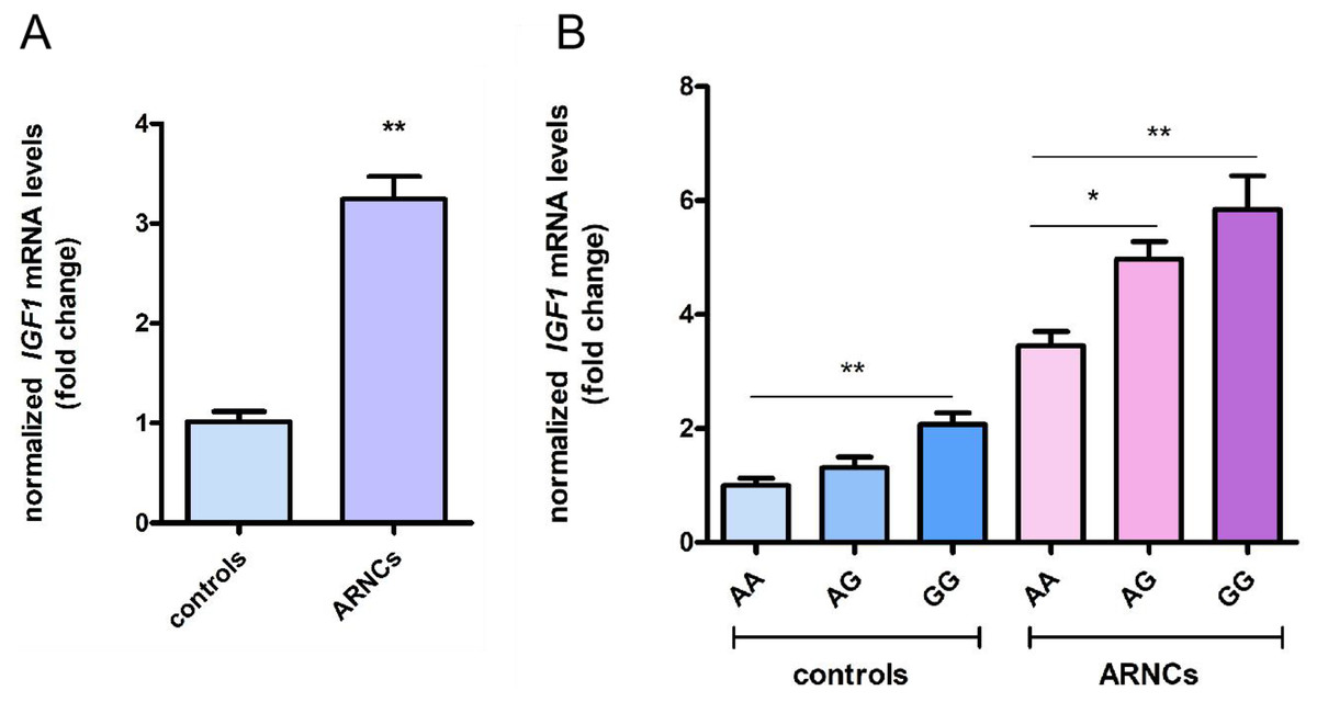
Figure 1: Levels of IGF1 mRNA expression of LECs in anterior capsules.
(A) IGF1 mRNA levels were higher in ARNCs than in the controls (20 ARNC patients and 20 age-, sex- and ethnically matched controls). (B) IGF1 mRNA levels were higher in the GG or AG group than the AA group in the ARNCs (AA genotype = 10; AG genotype = 4; GG genotype = 6). IGF1 mRNA levels were higher in the GG than the AA group in the controls (AA genotype = 10; AG genotype = 5; GG genotype = 5).*p < 0.05. **p < 0.01. The qRT-PCR were repeated three times independently. Data were presented as means ± SD.
Apoptosis of LECs in biopsy samples
Apoptosis was a normal physiological process that orderly controls cell death to maintain stable homeostasis. The experimental results showed that cell apoptosis was present in both the ARNCs and the control group of LECs. However, The apoptosis of LECs in the anterior capsules of ARNCs was significantly higher than in the controls (Fig. 2). *: p < 0.05.
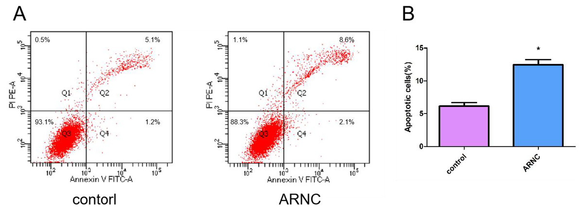
Figure 2: Representative images of cell apoptosis.
(A) FCM analysis of the effect on cell apoptosis in LECs in anterior capsules of controls and ARNCs (20 ARNC patients and 20 age-, sex- and ethnically matched controls). (B) Quantitative data of (A). *p < 0.05.
Discussion
Understanding the mechanism of ARC, which is recognized as the inaugural ocular disease causing blindness, is of utmost importance. SNPs contribute significantly to ARC development. Herein, Several genes and SNPs were revealed to be associated with ARCs (Cui et al., 2020; Kang et al., 2019; Zou et al., 2018, 2020).
LEC apoptosis has a close association with ARC formation, oxidative stress-induced apoptosis of LECs is the main factor in the pathogenesis of ARC (Tian et al., 2020; Zheng et al., 2018). Our previous research reported a correlation between oxidative damage of LECs and the occurrence and development of ARC (Kang et al., 2019; Zou et al., 2018), but did not detect apoptosis. Studies suggested that the LEC apoptosis level in ARC patients was significantly higher than in healthy individuals (Tian et al., 2020). In this study, we found the apoptosis of LECs in the anterior capsules of ARNCs was higher than that of the controls.
IGF system was found to be involved in the apoptosis regulation process (Willems et al., 2016; Tien et al., 2017). Wherein, IGF1 can inhibit cell proliferation and promote cell apoptosis (Cao et al., 2020). In addition, IGF1 has a close association with cataract formation (Civil et al., 2000; Papier et al., 2022; Wang et al., 2022). Study found IGF1R-rs1546713 may affect susceptibility to ARCs (Cui et al., 2020). In our study, we found the minor allele G frequency in the IGF1-rs6218 was higher in the ARCs than in the controls. Further performed stratification analysis revealed that the minor allele G frequency of IGF1-rs6218 was only higher in ARNCs than in the controls. This indicates that IGF1-rs6218 G allele might play a role in the formation and development of ARNC risk in Chinese population. This study also found that IGF1 mRNA expression was higher in the LECs of the ARNCs compared to the controls. Furthermore, the mRNA expression of IGF1 in GG-genotype individuals was higher than in AA-genotype individuals.
IGF1 can regulate cell apoptosis through targeted regulation by miRNA (Wang et al., 2020); rs6218 is located in the 3′-UTR region of the IGF1 gene that is predominantly related to the binding of microRNAs (miRNAs). The binding of miRNA to target genes leads to mRNA degradation or post-transcriptional inhibition, thereby inhibiting gene expression (Kang et al., 2019; Zheng et al., 2018). Consequently, we postulated that rs6218 might potentially alter the binding energy between IGF1 and miRNAs. Many miRNAs that can bind to IGF1 have been discovered, but unfortunately, no miRNAs that can bind near the rs6218 site have been found yet through the online database (https://compbio.uthsc.edu/miRSNP/miRSNP_detail_all.php). The aforementioned results implicated that there are additional underlying mechanisms behind IGF1 expression regulation in lens changing in LECs of ARNC patients. Nevertheless, this study had certain limitations; there are many reasons for the apoptosis of LECs, which may not be solely the result of IGF1 upregulation. Further experiments are needed to detect apoptosis by enhancing or knocking out the IGF1 gene in vitro experiments.
Conclusion
IGF1-rs6218 G allele might play a role in the formation and development of ARNC risk in Chinese population, and this polymorphism is associated with IGF1 expression at the mRNA level. IGF1 mRNA expression was higher in the LECs of the ARNCs compared to the controls. In addition, the expression of IGF1 mRNA in individuals carrying the G allele genotype was higher than that in individuals carrying the G allele genotype in both the ARNCs and controls. Moreover, apoptosis in LECs of ARNCs was found to be increased compared to the controls.
Supplemental Information
Raw data exported for the epidemiological participants.
Includes age (hidden), gender (1 is male, 2 is female), control or various types of ARC, as well as genotype of each candidate IGF1 gene locus.
Raw data exported for the hospitaI GF1l participants.
20 ARNC patients and 20 control patients. Raw data from qRT PCR of mRNA levels of IGF1 in lens anterior capsule. This also includes the genotypes of -rs2968 for these participants.