
Identification of a native Bacillus thuringiensis strain from Sri Lanka active against Dipel-resistant Plutella xylostella
- Published
- Accepted
- Received
- Academic Editor
- Brenda Oppert
- Subject Areas
- Agricultural Science, Entomology, Microbiology
- Keywords
- Bacillus thuringiensis, Plutella xylostella, Cry toxin, Resistance
- Copyright
- © 2019 Baragamaarachchi et al.
- Licence
- This is an open access article distributed under the terms of the Creative Commons Attribution License, which permits unrestricted use, distribution, reproduction and adaptation in any medium and for any purpose provided that it is properly attributed. For attribution, the original author(s), title, publication source (PeerJ) and either DOI or URL of the article must be cited.
- Cite this article
- 2019. Identification of a native Bacillus thuringiensis strain from Sri Lanka active against Dipel-resistant Plutella xylostella. PeerJ 7:e7535 https://doi.org/10.7717/peerj.7535
Abstract
Background
Biopesticides based on strains of the bacterium Bacillus thuringiensis (Bt) are used globally for effective and environmentally friendly pest control. The most serious threat to the sustainable use of these microbial pesticides is the development of resistance on targeted pests. Populations of Plutella xylostella (diamondback moth) have evolved field resistance to Bt pesticides at diverse locations worldwide. Discovery of novel Bt strains with varied toxin profiles that overcome resistance is one of the strategies to increase sustainability of Bt pesticides against P. xylostella. In this study, we report isolation and characterization of a Bt strain named AB1 from Sri Lanka displaying toxicity towards larvae of P. xylostella resistant to the commercial Bt pesticide Dipel.
Methods
Strains of Bt from diverse environments in Sri Lanka were evaluated for protein crystal production through Differential Interference Contrast (DIC) microscopic examination, and for insecticidal activity against P. xylostella in bioassays. The genome of the AB1 strain was sequenced by Hiseq Illumina sequencing to identify the insecticidal genes present in the genome and nano liquid chromatography followed by tandem mass spectrometry (nanoLC/MS/MS) of purified crystal proteins of AB1 was performed to identify the expressed insecticidal proteins. Multilocus sequence typing and Gyrase B gene sequence analyses were performed to identify the phylogenetic origin of the AB1 strain.
Results
The AB1 strain was identified as producing high levels of bipyramidal crystals and displaying insecticidal activity against susceptible and Dipel-resistant strains of P. xylostella. Multilocus sequence typing and phylogenetic analysis of the Gyrase B gene identified that AB1 belongs to the B. thuringiensis subsp. aizawai serotype. Comparative analysis of genomic and proteomic data showed that among the insecticidal protein coding genes annotated from the AB1 genome (cry1Aa, cry1Ca, cry1Da, cry1Ia, cry2Ab and cry9), Cry1Ca and Cry1Da toxins represented most of the toxin fraction in parasporal crystals from AB1. Overall findings warrant further development of B. thuringiensis subsp. aizawai AB1 strain as a pesticide to control P. xylostella.
Introduction
Strains of the bacterium Bacillus thuringiensis (Bt) produce a number of parasporal crystal proteins with insecticidal activity. Their high specificity and associated environmental safety have contributed to make Bt the most successfully commercialized biocontrol agent worldwide (Glare et al., 2012; Jurat-Fuentes & Crickmore, 2017). For example, the Dipel pesticide is based on B. thuringiensis subsp. kurstaki HD-1, which produces insecticidal proteins Cry1Aa, Cry1Ab, Cry1Ac, Cry2Aa and Cry2Ab as active ingredients (Höfte & Whiteley, 1989; Abbott Laboratories, 1992) to control lepidopteran larvae. The mode of action of these Cry toxins includes a number of successive steps, yet binding of the toxin to receptors on the midgut epithelium in the host is considered the most critical given that high levels of resistance are commonly linked to reduced toxin binding (Jurat-Fuentes & Crickmore, 2017). This resistance represents the main threat to the sustainability of Bt pesticides.
Plutella xylostella (Diamondback moth, Lepidoptera: Plutellidae) is a destructive agricultural pest attacking crucifer crops worldwide (Talekar & Shelton, 1993; Furlong, Wright & Dosdall, 2013) that is controlled by Bt pesticides. Annual global crucifer yield loss and control costs of P. xylostella together represent USD 2.7 billion (Zalucki et al., 2012; Furlong, Wright & Dosdall, 2013). Its high reproductive potential, voracious feeding, high genetic elasticity, and cosmopolitan distribution makes P. xylostella difficult to control (Machekano, Mvumi & Nyamukondiwa, 2017). Moreover, populations of P. xylostella have developed field-evolved resistance to virtually all used insecticides, including Bt pesticides such as Dipel (Kirsch & Schmutterer, 1988; Tabashnik et al., 1996; Heckel et al., 2001; Zhao et al., 2002; Furlong, Wright & Dosdall, 2013). Since resistance to Dipel was associated with reduced binding of Cry1A toxins (Ferre et al., 1991) it is expected that the use of B. thuringiensis strains or transgenic crops producing insecticidal proteins binding to distinct sites from those of Cry1A toxins would be effective against Dipel-resistant P. xylostella and assist in delaying resistance evolution (Zhao et al., 2003).
In this study, we report the identification of a B. thuringiensis strain from Sri Lankan soil displaying insecticidal activity against P. xylostella, including a Dipel-resistant strain (NO-QAGE). The sequenced genome of this strain was used as a reference database for proteomic identification of insecticidal proteins present in its parasporal body. The results support that toxicity of this strain against the NO-QAGE strain of P. xylostella is related to the production of Cry toxins not sharing binding sites with Cry1A toxins produced by B. thuringiensis subsp. kurstaki.
Materials & Methods
Bacterial strains and Plasmids
Reference strains B. thuringiensis subsp. kurstaki HD-1 (ATCC 33679) and B. thuringiensis subsp. israelensis (ATCC 35646) were purchased from the American Type Culture Collection (ATCC, Manassas, VA, USA). Strains, B. thuringiensis subsp. aizawai HD-133 (BGSC 4J1, 4J3) and B. thuringiensis subsp. tenebrionis (BGSC 4AA1) were purchased from the Bacillus Genetic Stock Center (BGSC, Ohio State University, Columbus, OH, USA). The TA cloning vector pGEM-T Easy used in this study was purchased from Promega (Madison, WI, USA).
Isolation and identification of Bacillus thuringiensis strains
Previously, B. thuringiensis strains were isolated from 63 soil samples from natural habitats in Sri Lanka (Travers, Martin & Reichelderfer, 1987). Isolates were designated with given numbers and are stored at the Industrial Technology Institute B. thuringiensis collection (Colombo, Sri Lanka). Isolated spore-forming Bacilli were grown on chromogenic Bacillus agar (HiMedia, Mumbai, India), to identify putative Bt colonies based on their blue–green color and irregular margins.
Putative Bt isolates were sub-cultured on Luria-Bertani (LB) agar plates and incubated at 30 ± 2 °C for 16 h to obtain pure cultures, which were used for genomic DNA extraction using the PureLink Genomic DNA Mini Kit (Invitrogen), following manufacturer’s instructions. The gyrase B (gyrB) gene was amplified from genomic DNA using nested PCR primers gyrB-F1 (5′ATGGAACAAAAGCAAATGCA-3′) and gyrB-R1 (5′TTAAATATCAAGGTTTTTCA-3′), and gyrB-F2 (5′-CCTTGYTTTGCWGAWCCDCC-3′) and gyrB-R2 (5′ACWCGTATGCGTGARYTRGC-3′) as second primer pair (Soufiane & Côté, 2009). The first PCR was carried out in 25 µL containing template genomic DNA (250 ng), 1× reaction buffer, 200 µM dNTPs, 1 mM MgCl2, 0.8 µM (each) primer and 1.25 U of Taq DNA polymerase (Promega). Amplification included initial denaturation at 95 °C for 3 min followed by 30 amplification cycles of denaturation at 95 °C for 30 s, annealing at 45 °C for 30 s and extension at 72 °C for 30 s in a Veriti thermocycler (Applied Biosystems, USA). A final extension step of 7 min at 72 °C was added after completion of 30 cycles. The second PCR was performed in a 50 µL reaction including 2 µL of the amplicon from the first PCR as template DNA, 1× reaction buffer, 200 µM dNTPs, 1 mM MgCl2, 0.8 µM (each) primer and 2.5 U of Taq DNA polymerase (Promega). Amplification included initial denaturation at 95 °C for 3 min followed by 30 amplification cycles of denaturation at 95 °C for 45 s, annealing at 50 °C for 30 s and extension at 72 °C for 1 min. A final extension step of 7 min at 72 °C was added after completion of 30 cycles. Amplified products were purified from 1% low-melting agarose using PureLink Quick Gel Extraction Kit (Invitrogen, Waltham, MA, USA) according to the manufacturer’s instructions. Purified amplicons of gyrB were bi-directionally sequenced using the Big Dye Terminator Cycle sequencing kit in an Applied Biosystems 3500 dx genetic analyzer (Applied Biosystems). Forward and reverse sequences were aligned and edited using the BioEdit sequence alignment editor (version 7.2.5). The sequences were used as query over the GenBank database at NCBI using BLAST searches.
Detection of insecticidal protein genes
Polymerase chain reaction (PCR) with specific primers was used to detect insecticidal genes (cry, vip and cyt) in isolated Bt strains. Strains were cultured in 10 mL of LB broth enriched with salts (0.2% NH4SO4, 1.4% K2HPO4, 0.6% KH2PO4, 0.1% sodium citrate, 0.02% MgSO4.7H2O) and 0.5% glucose at 30 °C, shaking at 200 rpm for 17 h, and then plasmid DNA was extracted (Reyes-Ramırez & Ibarra, 2008). Reference strains; B. thuringiensis subsp. kurstaki HD-1 (ATCC 33679), B. thuringiensis subsp. israelensis (ATCC 35646), B. thuringiensis subsp. aizawai HD-133 (BGSC 4J1, 4J3) and B. thuringiensis subsp. tenebrionis (BGSC 4AA1) served as controls in PCR reactions. Bacillus thuringiensis strains were screened for lepidopteran-specific cry and vip genes (cry1, cry2, cry9 and vip3A), Coleopteran-specific cry genes (cry3, cry7 and cry8), and Dipteran-specific cry and cyt genes (cry4, cry10, cry11, cyt1 and cyt2), using universal and gene specific primers (Ben-Dov et al., 1997; Ben-Dov et al., 1999; Bravo et al., 1998; Ejiofor & Johnson, 2002; Porcar & Juárez-Pérez, 2003). Each amplification reaction (25 µL) contained 200 µM dNTP, 0.5 µM of each primer, 2.5 mM MgCl2, and 2.5 U of Taq DNA polymerase. Amplifications were carried out in thermocycler following a program of initial denaturation at 94 °C for 3 min, followed by 35-cycles of denaturation at 94 °C for 45 s, annealing at corresponding annealing temperatures of the primer (Table S1) for 45 s and an extension at 72 °C for 1 min. A final extension step of 7 min at 72 °C was included after completion of 35 cycles (Ben-Dov et al., 1997; Bravo et al., 1998). Reaction products were resolved in 1% agarose gels and examined for the presence of the expected amplicon size.
Observation and purification of insecticidal crystal proteins (ICPs)
Bacillus thuringiensis strains were grown on 1/3 Tryptic Soy Broth (TSB) agar for 3 days at 28 °C until sporulation. Individual colonies were suspended in a drop of sterile distilled water and observed in a Differential Interference Contrast (DIC) microscope to document the presence of crystals.
For production of ICPs from B. thuringiensis strain AB1, three colonies of a sporulated culture were inoculated separately into 1 mL of sterile distilled H2O, vortexed to homogenize and heated at 70 °C for 45 min to kill vegetative cells and synchronize the culture. This pre-inoculum (500 µL) was used to inoculate 500 mL of 1/3 TSB, which were then incubated at 28 °C with shaking at 160 rpm for 3 days until 90% of cells appeared sporulated under microscopic inspection. Parasporal crystal proteins were isolated from spores using solubilization in 50 mM Na2CO3 containing 0.1% 2-mercaptoethanol, as described elsewhere (Perera et al., 2009). Solubilized crystal proteins were dialyzed overnight against 2 L Na2CO3 buffer (50 mM) with two changes of buffer, to eliminate 2-mercaptoethanol, and then stored at 4 °C until used (within 1 week).
Proteins present in the isolated parasporal crystal protein mixtures from strain AB1 were analyzed using electrophoresis. Aliquots (10 µL) of each protein sample were mixed with equal volume of 2% electrophoresis buffer (100 mM Tris pH 6.8, 4% SDS, 20% glycerol, 0.2% bromophenol blue, 2% β-mercaptoethanol), and heat-denatured for 10 min before loading on lanes of an SDS-10% PAGE gel. After the electrophoresis run, proteins were detected by staining with ProtoBlue Coomassie stain (National Diagnostics, Atlanta, GA, USA).
Insects and bioassays
Eggs of Dipel susceptible and resistant (NO-QAGE) strains of P. xylostella were purchased from Benzon Research (Carlisle, PA). The NO-QAGE strain was derived from a strain with field-evolved resistance to Dipel from Hawaii (Tabashnik et al., 1990), and it displays 500-1,000-fold resistance to B. thuringiensis subsp. kurstaki (https://www.benzonresearch.com/species-detail). Eggs were placed in an incubator (27 °C, 21% relative humidity, and 16-h light/8-h dark photoperiod) until hatched.
Insecticidal activity of isolated solubilized crystal proteins was evaluated first against Dipel-susceptible P. xylostella larvae using surface-diet contamination bioassays. Freshly prepared meridic diet (General Lepidopteran Diet; Frontier Agricultural Sciences, Newark, DE, USA) was dispensed into 128 well bioassay trays (CD International, Pitman, NJ) and allowed to solidify. Toxin (crystal protein) concentrations were measured using a Qubit 4 Fluorometer (Thermo Fisher, Waltham, MA, USA). Selected concentrations of solubilized crystal protein solutions (75 µL) were applied to each well ensuring uniform distribution. Crystal solubilization buffer (50 mM Na2CO3) without 2-mercaptoethanol was used as control treatment. After drying, five neonates were introduced into each well before sealing with adhesive tray lids (CD International, Pitman, NJ). Trays were incubated at 27 °C, 21% relative humidity, and 16-h light/8-h dark photoperiod and mortality recorded after 7 days. Bioassays were replicated three times with 20 larvae tested per each concentration. Further bioassays were performed with larvae from susceptible and Dipel-resistant (NO-QAGE) strains of P. xylostella to estimate median lethal concentration (LC50) of solubilized crystal proteins from the most active B. thuringiensis strains in bioassays above. These bioassays were conducted as above, except for testing eleven crystal protein concentrations against 50 larvae from each strain per concentration, and bioassays replicated three times with larvae from different cohorts. Mortality data were analyzed by probit analysis using PoloPlus 1.0 (PoloPlus, 1987) to estimate the LC50s and 95% fiducial limits. The ratio between LC50 of AB1 against the susceptible and Dipel-resistant P. xyslotella strains and approximate 95% confidential boundaries were estimated as described elsewhere (Robertson et al., 2007).
Genome sequencing and assembly
Genomic DNA of B. thuringiensis strain AB1 was purified as described above. DNA was quantified using Qubit 4 fluorometer (Thermo Fischer Scientific, Waltham, MA, USA) and randomly sheared using Covaris M220 Focused-ultrasonicator (Covaris, Woburn, MA, USA) to sizes between 200 and 500 bp per the manufacturer’s instructions (Barchenger et al., 2017). PCR free Illumina libraries were constructed using KAPA Hyper Prep Kit (Kapa Biosystems, Roche, Basel, Switzerland) followed by library quantification by qPCR KAPA Library Quantification Kit according to manufacturer’s instructions. The library was sequenced on Illumina Hiseq X at Admera Health (Illumina, San Diego, CA, USA). High throughput sequencing generated 38,737,140 high quality reads with an average length of 151 bp. The raw reads were deposited into Sequence Read Archive (SRA) database under NCBI accession PRJNA514414. Raw reads were quality trimmed and assembled (de novo) using CLC Genomic Workbench 11.0.1 software (Qiagen Bioinformatics, Redwood City, CA). De novo assembly of paired reads resulted in 213 contigs (N50 = 67 kbp). The assembled genome was submitted to the Prokaryotic GeneFinding tool in Blast2Go ver. 5.2.1 (Conesa et al., 2005) to obtain a FASTA file listing all possible Open Reading Frames (ORFs) in the contigs, which was used for proteomic analysis (see below). Homologous sequences to the predicted ORFs were searched using BLASTX in CloudBlast of Blast2Go software. GO terms were then assigned to each BLAST hit sequence by performing Mapping function followed by Annotation function, where the sequences were annotated using an annotation rule to the assigned GO’s using Blast2Go.
Typing of Bacillus thuringiensis strain AB1
Multilocus sequence typing (MLST) analysis was performed using the PubMLST Bacillus cereus database (https://pubmlst.org/bcereus/) to identify the sequence type (genetically similar subspecies) of strain AB1. The AB1 genome was queried to identify the seven sequence type loci (glpF, gmk, ilvD, pta, pur, pycA and tpi) used for MLST with B. thuringiensis strains (Wang et al., 2018). The PubMLST database was then queried to determine the allele pattern for the seven loci, which matched the profile of sequence type 15 (ST15).
The full-length GyrB gene sequence from strain AB1 was identified as contig 122 in the AB1 genome using a BLAST in CLC Genomics Workbench ver. 11 (Qiagen Bioinformatics, Redwood City, CA, USA). Contig 122 was used as query in a BLAST search against NCBInr, and the 60 best matching coding sequences were used for sequence alignment in CLC Genomics Workbench. A neighbor joining phylogenetic tree was constructed using the aligned sequences. Stability of each node was assessed by bootstrap analysis with 1,000 replicates, and only bootstrap values >90% were considered for branches in the graphical representation of the tree.
Proteomic identification of crystal proteins
Proteins in solubilized isolated parasporal crystal samples were identified and relatively quantified using SDS-10% PAGE and nano liquid chromatography followed by tandem mass spectrometry (nano LC/MS/MS). Solubilized crystal proteins (10 µg) were resolved by Bis-Tris SDS-10% PAGE, the gel stained with Coommassie blue, and the entire mobility region excised. The gel piece was then submitted to MS Bioworks LLC (Ann Arbor, MI) for nano LC/MS/MS. Trypsin digestion was performed by washing the gel piece with 25 mM ammonium bicarbonate followed by acetonitrile. Gel was reduced with 10 mM dithiothreitol at 60 °C followed by alkylation with 50 mM iodoacetamide at room temperature and digested with trypsin for 4 h at 37 °C. Trypsin activity was quenched with formic acid. The processed protein sample (supernatant) was analyzed by nano LC/MS/MS in a Waters NanoAcquity HPLC system interfaced to a LTQ Orbitrap Velos mass spectrometer (Thermo Fisher Scientific, Waltham, MA, USA). Peptides were loaded on a trapping column and eluted over a 75 µM analytical column at 350 nL/min; both columns were packed with Luna C18 resin (Phenomenex, Torrance, CA, USA). The mass spectrometer was operated in data-dependent mode, with MS and MS/MS performed in the Orbitrap at 70,000 FWHM resolution and 17,500 FWHM resolution, respectively. The fifteen most abundant ions were selected for MS/MS. Data were queried against a database including all the translated protein sequences from predicted open reading frames identified in the B. thuringiensis AB1 strain genome through the Prokaryotic GeneFinding tool in Blast2Go, as described above. Querying was performed using the Mascot software (Matrix Science, London) with carbamidomethyl (C) as fixed modification, and oxidation (M), acetyl (protein N-terminus), and deamidation (NQ) as variable modifications. Additional parameters included 10 ppm peptide mass tolerance, 0.02 Da fragment mass tolerance and two maximum missed cleavages. Mascot DAT files were parsed into the Scaffold ver. 4.8.8 (Proteome Software Inc., Portland, OR, USA) for validation based on peptide identification, filtering and to create a non-redundant list. Peptide identifications were accepted if they could be established at greater than 95.0% probability by the Scaffold Local FDR algorithm. Protein identifications were accepted if they could be established at greater than 99.0% probability and contained at least 2 identified peptides. Protein probabilities were assigned by the Protein Prophet algorithm (Nesvizhskii et al., 2003). Proteins that contained similar peptides and could not be differentiated based on MS/MS analysis alone were grouped to satisfy the principles of parsimony. The list of identified proteins was organized based on protein abundance considering the Normalized Spectral Abundance Factor (NASF) quantitative method (Zhang et al., 2010) which takes into account normalized spectra and protein length.
Results
Isolation and identification of Bacillus thuringiensis strains from Sri Lanka
Twenty-one B. thuringiensis strains were isolated from sixty-three soil samples collected from natural habitats in Sri Lanka (Table 1). For rapid and efficient isolation of Bt, we combined the conventional Travers method (Travers, Martin & Reichelderfer, 1987) with chromogenic identification of Bt as blue/green color colonies in Bacillus agar (HiChrome) (Fig. 1), making the identification of Bt from other spore-forming Bacilli much faster and easier.
| Bacillus thuringiensisstrain | GenBankgyrBgene accession number | Insecticidal gene profile | ICPs |
|---|---|---|---|
| AB1 | KF460446 | Cry 1, Cry4, Cry 9, Cry 10, Cry 11, Vip 3A | + |
| AB2 | KF481970.1 | Cry 2, Cry4, Cry 9, Cry 10, Cry11 | + |
| AB6 | KX758551 | Cry 1, Cry 9, Cry 10 | – |
| AB7 | KX758555 | Cry 1, Cry 9, Cry 10, Vip 3A | – |
| AB8 | KX758556 | Cry 1, Cry 10, Vip 3A, Cyt 1 | + |
| AB10 | KX758557 | Cry 1, Cry 9, Cry 10, Cyt 1 | – |
| AB11 | KX758553 | Cry 9, Cry 10 | – |
| AB12 | KX758558 | Cry 8, Cry 9, Cry 10 | – |
| AB13 | KX768535 | Cry 9, Cry 10 | – |
| AB14 | KX768536 | Cry 1, Cry 8, Cry 10, Cyt 1 | – |
| AB15 | KX758552 | Cry 1, Cry 2, Cry 8, Vip 3A | + |
| AB16 | KX758554 | Cry 1, Cry 2, Vip 3A | – |
| AB17 | KX758548 | Cry 1, Cry 2, Cry 9, Cry 10, Vip 3A | – |
| AB19 | KX758549 | Cry 9, Vip 3A | – |
| AB20 | KX758545 | Cry 8 | + |
| AB21 | KX758547 | Cry 9, Cry 10 | + |
| AB22 | KX758544 | Cry 1, Cry 2, Cry 9, Vip 3A, Cyt 2 | + |
| AB23 | KX758546 | Cry 9, Cry 10, Cyt 1 | – |
| AB24 | KX758550 | Cry 9, Cry 10, Cyt 1 | + |
| AB125 | KF444206 | Cry 2, Cry 4, Cry 9, Cry 10, Cry 11 | + |
| AB142 | KF444205 | Cry 1, Cry 4, Cry 10, Cry 11, Vip 3A | + |
Sequencing of the gyrB gene further identified the strains as Bt. Amplified gyrB sequences aligned with the gyrB gene sequences of reported B. thuringiensis strains available in the GenBank database with 99–100% sequence similarity. Sequence data have been deposited in GenBank (Table 1). The gyrB sequence data showed that the most common subspecies among the 21 tested Bt strains were graciosiensis (eight strains), kurstaki (five), konkukian (two) and israelensis (two).
Detection of insecticidal genes by PCR was used to predict insecticidal activity of native B. thuringiensis strains (Carozzi et al., 1991). Analysis of the 21 B. thuringiensis strains revealed the presence of amplified fragments characteristic of cry1, cry2, cry4, cry8, cry9, cry10, cry11, vip3A, cyt1 and cyt2 genes (Table 1). The most abundant (76% of strains) insecticidal gene among the screened B. thuringiensis strains was the dipteran-specific cry10. Among the lepidopteran-specific genes, cry1, cry2 and cry9 genes were present in 52%, 28% and 71% of the tested strains, respectively, while vip3A gene was present in 42% of the strains. The frequency of dipteran-specific cry4/cry11, cyt1 and cyt2 genes was 19%, 23% and 4%, respectively. Nineteen percent of strains harbored the coleopteran-specific cry8 gene.
In contrast to detection of ICP genes in all tested strains, not all B. thuringiensis strains produced observable parasporal crystal proteins (Table 1). In fact, only 5 (AB1, AB2, AB8, AB125, AB142) out of the 21 strains showed high amounts of clear crystal inclusions. Lower levels of crystals inclusions were observed in strains AB21, AB22 and AB24, while crystals were not detected in the remaining strains. Different morphologies, including rhomboidal, spherical and spore-attached crystals were observed (Fig. 2).
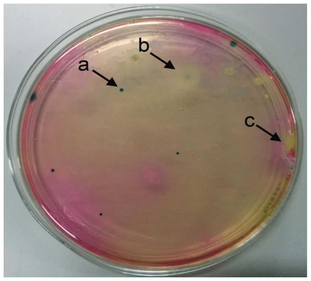
Figure 1: Selective identification of Bacillus thuringiensis colonies on Bacillus agar based on colony color.
Selective identification of Bacillus thuringiensis colonies on Bacillus agar. Bacillus thuringiensis colonies were grown as blue/green in color (A), Bacillus cereus appeared as white color colonies with a light blue center (B), and Bacillus megaterium colonies appeared yellow (C).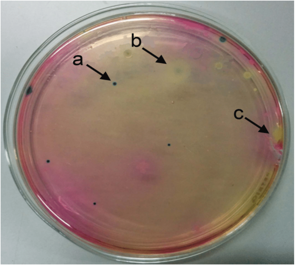
Toxicity of Bacillus thuringiensis strains against Plutella xylostella
Due to limited resources, we focused toxicity assays on B. thuringiensis strains classified as producing high amounts of crystals (AB1, AB2, AB8, AB125 and AB142). The composition of the solubilized crystal protein fractions was assessed by electrophoresis (Fig. 3). Diversity in protein profiles were observed among the tested strains, in agreement with the diverse toxin genes detected by PCR in each strain.
Figure 2: Differential Interference Contrast micrographs of Bacillus thuringiensis strains showing different morphologies of insecticidal crystal proteins (ICPs).
Differential Interference Contrast micrographs of Bacillus thuringiensis strains showing rhomboidal parasporal crystals found in strain AB1 (A) and spherical crystals observed in strain AB21 (B). Black arrows point to released crystals, white arrows point to spores.
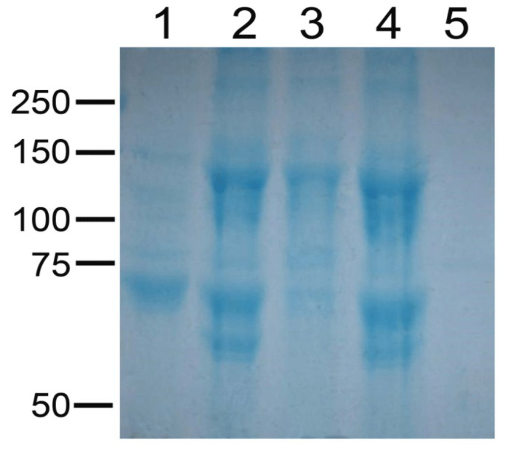
Figure 3: Electrophoretic pattern of solubilized crystal protein fractions from selected strains of Bacillus thuringiensis.
Electrophoretic pattern of solubilized crystal protein fractions of Bacillus thuringiensis strain AB1 (lane 1), Bacillus thuringiensis strain AB2 (lane 2), Bacillus thuringiensis strain AB8 (lane 3), Bacillus thuringiensis strain AB125 (lane 4) and Bacillus thuringiensis strain AB142 (lane 5).
Toxicity of the solubilized crystal protein fractions was assessed using a range of protein concentrations (from 0.1 µg to 10 µg /cm2) against larvae from Dipel susceptible and resistant populations of P. xylostella (Table 2). Percentage mortality data from this preliminary screen identified solubilized crystal proteins from B. thuringiensis strain AB1 as the most active against both susceptible and Dipel-resistant (NO-QAGE) strains of P. xylostella. Solubilized crystal proteins from strains AB2, AB8 and AB125 displayed similar toxicity against susceptible P. xylostella compared to AB1, but they did not present comparable toxicity against larvae from the NO-QAGE strain. B. thuringiensis strain AB142 displayed the lowest levels of activity against both strains of P. xylostella tested (Fig. 4).
Figure 4: Toxic effect of isolated crystal proteins from Bacillus thuringiensis strains to neonates of Dipel-resistant-DR (NO-QAGE) and susceptible DS Plutella xylostella strains.
Data represent mean mortality (%) ± SEM of 3 independent experiments.
Characterization of Bacillus thuringiensis strain AB1
The AB1 strain of B. thuringiensis was selected for further characterization based on its relatively higher toxicity against larvae from the susceptible and NO-QAGE strains of P. xylostella. This strain was deposited and is archived in the B. thuringiensis strain collection at the Industrial Technology Institute in Colombo (Sri Lanka), with accession number ITI-SL-AB1-2013. The genome of the strain was sequenced using Hiseq Illumina to identify insecticidal protein genes present and to generate a custom database for proteomic querying in the identification of insecticidal proteins among the solubilized crystal proteins.
Concentration-mortality assays with the solubilized crystal proteins from B. thuringiensis strain AB1 were performed to estimate toxicity parameters (Table 2). While not statistically significant due to overlapping fiducial limits values (Gonzalez-Cabrera et al., 2001; Mushtaq, Shakoori & Jurat-Fuentes, 2018), results from these bioassays identified a lower LC50 for these proteins against larvae from the NO-QAGE (0.091 µg/cm2) compared to the susceptible (0.313 µg/cm2) strain. The ratio of LC50 values and the corresponding 95% confidence intervals calculated as described by Robertson et al. (2007) suggested a 3.4-fold difference in susceptibility between the strains with 2.0 and 5.8 as lower and upper limits.
We used high throughput sequencing to further identify genes expressed in B. thuringiensis strain AB1. Assembly of reads resulted in 213 contigs with and N50 of 67 kb. Genome annotation results showed that B. thuringiensis strain AB1 contained pesticidal genes cry1Aa, cry1Ca, cry1Da, cry1Ia, cry2Ab, cry9Ea and vip3A.
Nano LC/MS/MS was performed on the solubilized crystal proteins of strain AB1 to identify the insecticidal genes expressed in the strain. Queries of MS data against the database of predicted ORFs from the genome confirmed the presence of 5 insecticidal Cry proteins in the AB1 sample (Table 3). Among these detected Cry toxins, Cry1Ca and Cry1Da were the most abundant, representing 61.2% and 31.2% of the total normalized spectra. The other three toxins detected, Cry1Aa, Cry1Ia, and Cry2Ab, represented <3% of the normalized spectra (Table 3).
| Identified protein | Accession number | Description | EUPCa | NSAFb |
|---|---|---|---|---|
| Cry1Ca | WP_042991625.1 | Pesticidal protein | 120 | 96.6 |
| Cry1Da | WP_042991627.1 | Pesticidal crystal protein Cry1Da | 70 | 19.6 |
| Cry1Aa | WP_00369823.1 | Pesticidal crystal protein Cry1Aa | 6 | 0.671 |
| Cry1Ia | WP_000769223.1 | Pesticidal crystal protein Cry1Ia | 3 | 0.471 |
| Cry2Ab | WP_001089638.1 | Pesticidal crystal protein Cry2Ab | 3 | 0.402 |
Typing of Bacillus thuringiensis strain AB1
Multilocus sequence typing using the AB1 genome in the PubMLST database identified the allele pattern for the seven loci used for classification: nine (glp), eight (gmk), 16 (ilv), 13 (pta), two (pur), 16 (pyc A) and nine (tpi), which matched the profile of sequence type 15 (ST15). This ST15 group comprises 12 bacterial isolates, of which eight are Bt. Seven out of the eight Bt isolates belong to serovar aizawai, supporting that strain AB1 belongs to this serovar.
Further evidence for serotyping of strain AB1 as aizawai was obtained through analysis of the full length gyrB gene sequence. A neighbor-joining tree constructed using the gyrB sequence from the strain AB1 genome (contig 122) and the 60 most similar gyrB sequences in the NCBInr database (Fig. 5), identified different clades (only considering branches with bootstrap values >90%). Strain AB1 was clustered in a clade together with strains of Bacillus thuringiensis serovar aizawai.
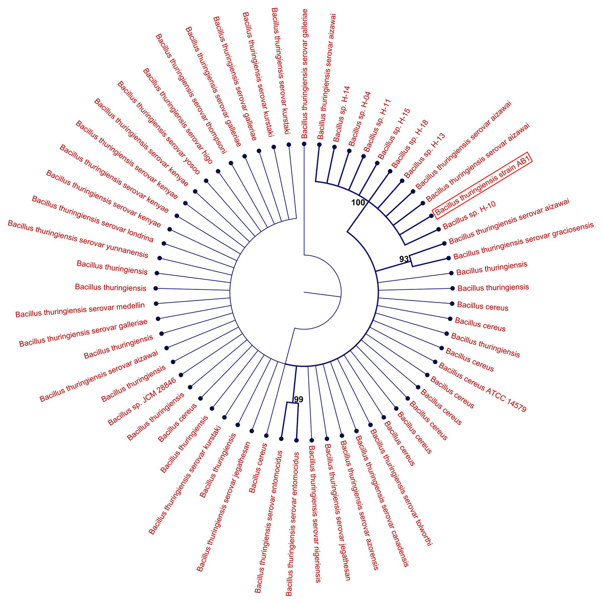
Figure 5: Neighbor-joining tree displaying identity in the gyrB gene sequence between the AB1 strain and the 60 most closely identical gyrB gene sequences in the NCBInr database.
Stability of each node was assessed by bootstrap analysis with 1,000 replicates, and only bootstrap values >90% were considered for branches in the graphical representation of the tree.
Discussion
Biopesticides based on the bacterium Bt account for about 80% of the microbial pesticide market and represent a safer and more environmentally friendly approach alternative to synthetic pesticides, which display broad toxicity. These Bt pesticides have been used for decades for the control of P. xylostella larvae, the most devastating pest of crucifer crops worldwide (Sarfraz, Keddie & Dosdall, 2005). Intensive use of Bt pesticides to control P. xylostella has resulted in evolution of resistance in populations at diverse locations worldwide (Tabashnik et al., 1996; Heckel et al., 2001) highlighting the importance of discovering new B. thuringiensis strains that overcome resistance. In this regard, we report identification of B. thuringiensis strain AB1 isolated from Sri Lankan soil as toxic against susceptible and Dipel-resistant strains of P. xylostella.
Among 21 Bt strains characterized, strain AB1 displayed the highest relative levels of activity against P. xylostella. Interestingly, strains AB2 and AB8 displayed similar levels of activity against susceptible P. xylostella but were not as active against the NO-QAGE larvae. Based on these observations, we limited detailed characterization to strain AB1.
Both MLST and analysis of the full length gyrB gene typed strain AB1 as B. thuringiensis subsp. aizawai. In agreement with this conclusion, analysis of the flagellin gene showed highest similarity of AB1 to B. thuringiensis subsp. aizawai (ETE87447). In addition, the insecticidal gene composition of strain AB1 mostly resembles B. thuringiensis subsp. aizawai HD-133 (Jeong, Choi & Park, 2017). All these observations strongly support typing of strain AB1 as B. thuringiensis serovar aizawai.
Comparison of genomic and proteomic results support that the AB1 strain produces Cry1Aa, Cry1Ca, Cry1Da, Cry1Ia, and Cry2Ab proteins, albeit at diverse levels. The high level of toxicity of crystal proteins from strain AB1 against larvae from susceptible and NO-QAGE strains of P. xylostella is explained by the insecticidal crystal proteins produced by this strain. Proteomic data supported that Cry1C and Cry1D proteins make up to approximately 92% of the insecticidal crystal proteins in AB1. Neither of these toxins share binding sites with Cry1Ac in P. xylostella or any other lepidopteran larvae (Jakka, Ferré & Jurat-Fuentes, 2015), and they do not display cross-resistance in Cry1C- and Cry1D-resistant P. xylostella (Sayyed & Wright, 2001). Thus, “Mode 1” resistance in NO-QAGE, characterized for high resistance levels to at least one Cry1A protein and low or nil cross-resistance to Cry1C, is overcome by Cry1Ca and Cry1D in parasporal crystals from strain AB1. Attempts to sequence the cry1C gene in AB1 strain confirmed that it is >99% identical to the cry1Ca gene—JN651493.1 (data not shown).
Trace levels of Cry2Ab and Cry1I proteins were detected (three unique peptides matching to each) in the parasporal crystal proteome of strain AB1. Genes in both the Cry2Ab and Cry1I families are generally cryptic in nature (Tailor et al., 1992; Crickmore, Wheeler & Ellar, 1994), which probably explains the trace amounts detected for both proteins.
Conclusions
Overall, findings of this study identify a native Sri Lankan strain of B. thuringiensis (AB1) typed as serovar aizawai producing mostly Cry1Ca and Cry1D toxins in its parasporal crystals. Data from this study support the potential use of this new B. thuringiensis subsp. aizawai strain AB1 to control P. xylostella larvae and to delay evolution of resistance to Bt pesticides that rely on Cry1A toxicity for their efficacy. Further work will be needed to determine the feasibility of culturing B. thuringiensis subsp. aizawai strain AB1 for commercialization.